
IGF-1 LR3 vs IGF-1: What the Long R3 Changes Actually Do
For laboratory research only. Not for human or animal use. This article gives no dosing, administration or therapeutic advice.
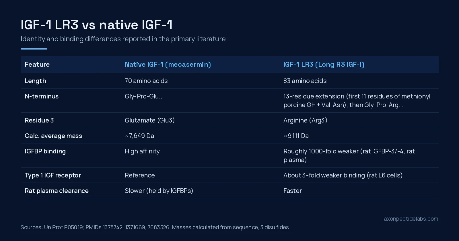
Short answer: IGF-1 LR3 (Long R3 IGF-I, often written LR3IGF-I) is an engineered version of human insulin-like growth factor-I. It carries two changes: a 13-residue N-terminal extension and an arginine in place of the glutamate at position 3 of the IGF-1 sequence[4][5]. Native IGF-1 is the unmodified 70-residue hormone, made as a recombinant drug under the name mecasermin[15]. The changes do not make LR3 bind the IGF-1 receptor better. In rat L6 myoblasts it bound the type 1 receptor about 3-fold less well than IGF-1[5]. What changes is its contact with the six IGF-binding proteins (IGFBPs): IGF-1 had roughly 1000-fold higher affinity for IGFBP-3, IGFBP-4 and total rat plasma binding proteins than LR3 did[6]. Almost every difference reported between the two in cells and animals traces back to that one property.
A note on what we sell: Axon lists IGF-1 LR3 1mg as a research peptide. We do not sell native IGF-1 or mecasermin. This article compares the two because the literature on LR3 was built by measuring it against native IGF-1, side by side.
Two molecules, one core sequence
Mature human IGF-1 is a single 70-residue chain with three disulfide bonds. Its sequence, residues 49 to 118 of the UniProt precursor record P05019, is GPETLCGAELVDALQFVCGDRGFYFNKPTGYGSSSRRAPQTGIVDECCFRSCDLRRLEMYCAPLKPAKSA. Its first three residues, Gly-Pro-Glu, turned out to matter far more than their size suggests.
In 1989 a group chemically synthesised IGF-1 analogues missing one to five residues from the N-terminus and tested them on rat L6 myoblasts. Removing the first three residues, giving des(1-3)IGF-1, made the peptide markedly more potent at stimulating protein synthesis, even though its binding to receptors on the cells barely changed. Cutting a fifth residue destroyed activity. The authors’ receptor-binding data also hinted that the myoblasts secreted a binding protein that held native IGF-1 but not the truncated form[1]. A second 1989 study made that explicit. A binding protein purified from bovine kidney cells strongly inhibited IGF-2 and partly inhibited IGF-1 in chick embryo fibroblasts, yet it did not affect des(1-3)IGF-1[2].
That gave the field a design rule: changes at the N-terminus can switch off binding-protein capture while leaving the receptor-binding core alone. LR3 was built on that rule.
How LR3 was built
The Adelaide group behind these papers first expressed IGF-1 in Escherichia coli as a fusion protein, with part of methionyl porcine growth hormone attached to the front of IGF-1 through a Val-Asn linker. They used the same system to make [Gly3]-IGF-I and [Arg3]-IGF-I, with Glu3 swapped for glycine or arginine. Both analogues were more potent than IGF-1 in L6 myoblast assays of protein and DNA synthesis. They bound bovine IGFBP-2 very poorly and the type 1 receptor slightly less well than IGF-1. The authors concluded that weaker binding-protein capture, not stronger receptor binding, explained the extra potency[3].
The next step kept a short piece of the fusion partner on the finished peptide. In the “Long” analogues, IGF-1 carries the first 11 amino acids of methionyl porcine growth hormone plus Val-Asn in front of its own sequence. Long IGF-I keeps the native sequence after that extension, and Long [Arg3]-IGF-I adds the Arg3 swap[4]. In cell lines that secrete IGFBPs into the medium, the potency order was Long [Arg3]-IGF-I and des(1-3)IGF-I highest, then Long [Gly3]-IGF-I, then Long IGF-I, then IGF-1. In chick embryo fibroblasts, which secrete no detectable IGFBPs, Long [Arg3]-IGF-I was less potent than IGF-1[4]. That control is the key result. Take away the binding proteins and the “more potent” analogue stops looking more potent.
Identity table
Neither molecule has a PubChem compound record (CID): PubChem returns no CID for “mecasermin”, “IGF-1” or “Long R3 IGF-1”, as is usual for proteins of this size. The values below come from the UniProt sequence record and the primary papers. The masses are our own calculation from the sequences (average isotopic masses, three disulfide bonds), not certificate-of-analysis values.
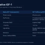
| Field | Native IGF-1 | IGF-1 LR3 |
|---|---|---|
| Other names | Insulin-like growth factor-I; recombinant form: mecasermin (rhIGF-1) | Long R3 IGF-I, LR3IGF-I, Long [Arg3]-IGF-I |
| Public identifier | UniProt P05019 (mature chain = residues 49-118); no PubChem CID | No PubChem CID or UniProt entry of its own (engineered analogue) |
| Length | 70 residues | 83 residues (13 + 70) |
| N-terminal sequence | GPE TLCGAE… | MFPAMPLSSLFVN GPR TLCGAE… |
| Position 3 of the IGF-1 sequence | Glu | Arg |
| Disulfide bonds | 3 | 3 (same core) |
| Average mass (calculated) | about 7,649 Da | about 9,111 Da |
| Source of design | Native hormone | Fusion-protein expression work, 1992[4] |
The LR3 sequence in the table is the 11 residues of methionyl porcine growth hormone (Met-Phe-Pro-Ala-Met-Pro-Leu-Ser-Ser-Leu-Phe), the Val-Asn linker, and then the IGF-1 sequence with Arg at position 3. On a mass spectrum the two differ by roughly 1,460 Da, so a lab can tell them apart without sequencing.
Side-by-side: what the papers measured
| Property | Native IGF-1 | IGF-1 LR3 | Model | Source |
|---|---|---|---|---|
| Affinity for IGFBP-3, IGFBP-4, rat plasma IGFBPs | About 1000-fold higher | Very low | Rat proteins, in vitro | [6] |
| Type 1 IGF receptor binding | Reference | About 3-fold weaker | Rat L6 myoblasts | [5] |
| Potency in IGFBP-secreting cells | Reference | 5- to 10-fold greater | Rat L6 myoblasts | [6] |
| Potency where no IGFBPs are secreted | Reference | Lower than IGF-1 | Chick embryo fibroblasts | [4] |
| Plasma clearance | Slower | Much faster | Rat | [6] |
| Binding in plasma across species | Bound in all species tested | Weakest in rat; binds significantly in sheep, human, pig, chicken plasma | Plasma, in vitro | [7] |
| Acute glucose lowering | Reference | 2- to 3-fold more potent and longer-lasting | Pig | [9] |
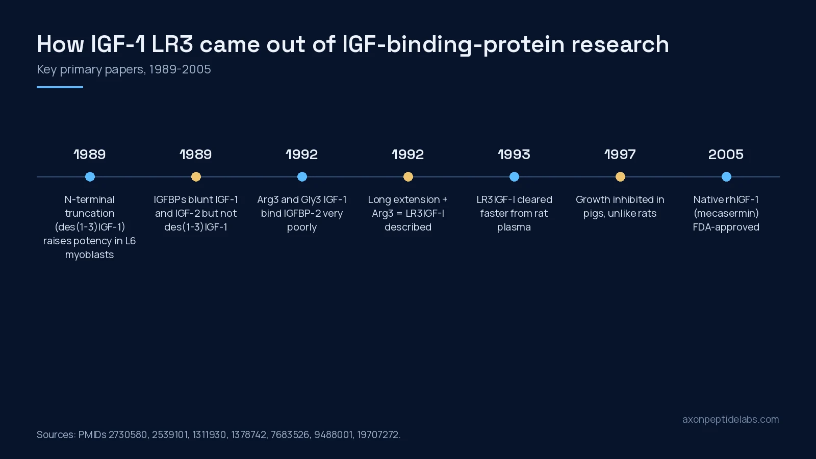
The binding-protein story, in more detail
IGFBPs are not just carriers. By holding IGFs away from the type 1 receptor they can block mitogenesis, differentiation and survival signals. Proteolysis of an IGFBP can release the IGF again, and some IGFBPs act on cells without IGF at all. IGFBP-1, for example, interacts with an integrin, and IGFBP-3 and -5 bind cell-surface and matrix proteins[14]. So any comparison between LR3 and IGF-1 in a system that contains IGFBPs is really a comparison of “IGF-1 plus whatever the binding proteins do to it” against “a ligand the binding proteins mostly ignore”.
A 2004 study shows why that matters in culture. Recombinant porcine IGFBP-3 suppressed proliferation of L6 myogenic cells driven by either IGF-1 or LR3, which the authors read as partly an IGF-independent effect. But IGFBP-3 suppressed IGF-1-driven differentiation of the same cells and did not suppress LR3-driven differentiation[12]. A cultured follicle study points the same way. Bovine preantral follicles grown in serum-free medium with LR3 or recombinant human IGF-1 both grew in diameter. However, LR3, and IGF-1 only at its highest concentration, gave smaller oocyte-to-follicle ratios and more oocyte degeneration. The authors concluded that IGFBP control of IGF availability is needed during early follicle development[13].
For a lab, the practical reading is that LR3 is a tool for asking what happens when IGF signalling escapes binding-protein control. It is not a drop-in “stronger IGF-1”.
What happened in animals
The rat work came first. In dexamethasone-treated rats, LR3IGF-I and des(1-3)IGF-I were about 2.5-fold more potent than IGF-1 at countering the catabolic state. The authors noted this was especially striking for LR3, which binds the receptor less well[5]. In normal female rats, LR3 was about 6-fold more potent than IGF-1 in growth measures over 14 days. It was also cleared from plasma much faster. In pregnant rats, where IGFBP levels are low, IGF-1 was cleared almost as fast as LR3, which fits binding-protein capture as the explanation[6]. Potency was partly kept when continuous infusion was replaced with injections, though continuous infusion worked better for both peptides[8].
The species results are where the simple story breaks down. In guinea pigs, which carry substantial IGF-1 and IGF-2 in their blood, a week of LR3 infusion increased the weights of some organs but did not stimulate overall growth. It also lowered plasma IGF-1, IGF-2 and IGFBP concentrations[10]. In finisher pigs, LR3 infusion reduced growth rate, food intake, plasma IGFBP-3, IGF-1 and insulin, and blunted growth hormone peaks. The authors note that analogues which stimulate growth in rats inhibit it in pigs[11]. In pigs and marmosets, IGF-1 variants that bind IGFBPs poorly lowered plasma glucose 2- to 3-fold more potently than IGF-1, and the effect lasted much longer[9]. The 1994 plasma study gives part of the reason: the variants that barely bind rat plasma proteins bind significantly in plasma from sheep, humans, pigs and chickens[7].
All of these are animal or cell studies from roughly 1989 to 2007. None of them is a human study of LR3.
Where native IGF-1 stands as a drug
Recombinant human IGF-1, mecasermin, has a regulatory history that LR3 does not. A 2009 review states that mecasermin was approved by the US Food and Drug Administration in 2005 and by the European Medicines Agency in 2007. It was approved for severe primary IGF-1 deficiency, a condition in which the growth hormone receptor or the IGF-1 gene is altered[15]. We report that status only as context. This article makes no claim about clinical use, and IGF-1 LR3 has no such approval.
What the evidence does not show
- No human data on LR3. We found no published human study of IGF-1 LR3. Everything above comes from cell lines, explants, rats, guinea pigs, pigs and marmosets.
- “More potent” depends on the system. The extra potency appears where IGFBPs are present. It disappears, or reverses, in cells without them[4], and growth effects in rats did not carry over to guinea pigs or pigs[10][11].
- “Longer-acting” is not a fact about plasma half-life. Competitor pages often give LR3 a long circulating half-life. In rats, the primary data show the opposite: LR3 was removed from plasma much faster than IGF-1[6]. Longer-lasting effects such as the glucose lowering in pigs are a separate finding[9].
- Species matters for binding. Rat plasma is the extreme case for weak LR3 binding, and results there do not transfer neatly to other species[7].
- Batch identity is a separate question. None of these papers says anything about a commercial vial. Identity and purity have to come from a certificate for the specific lot.
Documentation for our IGF-1 LR3
We checked our COA page and the product page while preparing this article. No certificate is posted for this product on our COA page at the time of writing. If a report is added later, it will be listed there with its batch number, purity result and test date. Related products sit in our growth hormone peptides category. For how LR3 differs from somatropin itself, see IGF-1 LR3 vs HGH. Researchers ordering supplies can buy lab-tested peptides in Canada from our main catalogue.
Frequently asked questions
What is the difference between IGF-1 LR3 and IGF-1?
IGF-1 LR3 is human IGF-1 with a 13-residue N-terminal extension and arginine instead of glutamate at position 3, giving 83 residues instead of 70. Those changes cut its binding to IGF-binding proteins about 1000-fold in rat studies, while its binding to the type 1 IGF receptor is about 3-fold weaker than IGF-1’s.
Why is IGF-1 LR3 more potent than IGF-1 in cell studies?
Because cells and plasma contain IGF-binding proteins that capture native IGF-1. LR3 largely escapes them, so more of it reaches the receptor. In chick embryo fibroblasts, which secrete no detectable binding proteins, Long [Arg3]-IGF-I was actually less potent than IGF-1.
Does IGF-1 LR3 have a longer half-life than IGF-1?
Not in plasma, in the study that measured it. In rats LR3 was cleared from plasma much faster than IGF-1, because binding proteins normally hold IGF-1 in circulation. Some effects of IGFBP-evading variants, such as glucose lowering in pigs, lasted longer, but that is a different measurement.
Is IGF-1 LR3 the same as mecasermin?
No. Mecasermin is recombinant native human IGF-1, approved by the FDA in 2005 and the EMA in 2007 for severe primary IGF-1 deficiency. IGF-1 LR3 is an engineered research analogue with no such approval. Axon sells IGF-1 LR3 for research only and does not sell mecasermin or native IGF-1.
Does IGF-1 LR3 have a PubChem CID?
No. Neither LR3 nor native IGF-1 has a PubChem compound record, which is normal for proteins of this size. Native IGF-1 is identified by UniProt P05019. LR3’s sequence and design come from the 1992 papers that described it.
- Bagley CJ, May BL, Szabo L et al. (1989). A key functional role for the insulin-like growth factor 1 N-terminal pentapeptide. Biochem J. PMID: 2730580. doi: 10.1042/bj2590665.
- Ross M, Francis GL, Szabo L et al. (1989). Insulin-like growth factor (IGF)-binding proteins inhibit the biological activities of IGF-1 and IGF-2 but not des-(1-3)-IGF-1. Biochem J. PMID: 2539101. doi: 10.1042/bj2580267.
- King R, Wells JR, Krieg P et al. (1992). Production and characterization of recombinant insulin-like growth factor-I (IGF-I) and potent analogues of IGF-I, with Gly or Arg substituted for Glu3, following their expression in Escherichia coli as fusion proteins. J Mol Endocrinol. PMID: 1311930. doi: 10.1677/jme.0.0080029.
- Francis GL, Ross M, Ballard FJ et al. (1992). Novel recombinant fusion protein analogues of insulin-like growth factor (IGF)-I indicate the relative importance of IGF-binding protein and receptor binding for enhanced biological potency. J Mol Endocrinol. PMID: 1378742. doi: 10.1677/jme.0.0080213.
- Tomas FM, Knowles SE, Owens PC et al. (1992). Insulin-like growth factor-I (IGF-I) and especially IGF-I variants are anabolic in dexamethasone-treated rats. Biochem J. PMID: 1371669. doi: 10.1042/bj2820091.
- Ballard FJ, Walton PE, Bastian S et al. (1993). Effects of interactions between IGFBPs and IGFs on the plasma clearance and in vivo biological activities of IGFs and IGF analogs. Growth Regul. PMID: 7683526.
- Lord AP, Bastian SE, Read LC et al. (1994). Differences in the association of insulin-like growth factor-I (IGF-I) and IGF-I variants with rat, sheep, pig, human and chicken plasma-binding proteins. J Endocrinol. PMID: 7514204. doi: 10.1677/joe.0.1400475.
- Tomas FM, Lemmey AB, Read LC et al. (1996). Superior potency of infused IGF-I analogues which bind poorly to IGF-binding proteins is maintained when administered by injection. J Endocrinol. PMID: 8708565. doi: 10.1677/joe.0.1500077.
- Tomas FM, Walton PE, Dunshea FR et al. (1997). IGF-I variants which bind poorly to IGF-binding proteins show more potent and prolonged hypoglycaemic action than native IGF-I in pigs and marmoset monkeys. J Endocrinol. PMID: 9415072. doi: 10.1677/joe.0.1550377.
- Conlon MA, Tomas FM, Owens PC et al. (1995). Long R3 insulin-like growth factor-I (IGF-I) infusion stimulates organ growth but reduces plasma IGF-I, IGF-II and IGF binding protein concentrations in the guinea pig. J Endocrinol. PMID: 7561636. doi: 10.1677/joe.0.1460247.
- Dunaiski V, Dunshea FR, Walton PE et al. (1997). Long [R3] insulin-like growth factor-I reduces growth, plasma growth hormone, IGF binding protein-3 and endogenous IGF-I concentrations in pigs. J Endocrinol. PMID: 9488001. doi: 10.1677/joe.0.1550559.
- Xi G, Kamanga-Sollo E, Pampusch MS et al. (2004). Effect of recombinant porcine IGFBP-3 on IGF-I and long-R3-IGF-I-stimulated proliferation and differentiation of L6 myogenic cells. J Cell Physiol. PMID: 15254966. doi: 10.1002/jcp.20068.
- Thomas FH, Campbell BK, Armstrong DG et al. (2007). Effects of IGF-I bioavailability on bovine preantral follicular development in vitro. Reproduction. PMID: 17636166. doi: 10.1530/REP-06-0382.
- Firth SM, Baxter RC (2002). Cellular actions of the insulin-like growth factor binding proteins. Endocr Rev. PMID: 12466191. doi: 10.1210/er.2001-0033.
- Fintini D, Brufani C, Cappa M (2009). Profile of mecasermin for the long-term treatment of growth failure in children and adolescents with severe primary IGF-1 deficiency. Ther Clin Risk Manag. PMID: 19707272. doi: 10.2147/tcrm.s6178.